2011年4月24日星期日

light reaction

      Photosynthesis can be divided into two processes: the light reaction and carbon fixation.

Light reaction:
Photoexcitation: absorption of a photon by an electron of chlorophyll.
      During the interaction with a photon, the electron gains energy and is raised to a higher potential energy level. This process is called excitation. However, an excited electron is unstable, and it will return to its ground state again. The loss in potential energy appears as heat and light. This process is called fluorescence. In chlorophyll, since it is normally embedded in the photosynthetic membrane, it will not undergo fluorescence but the excited electron is captured by a primary electron acceptor in the photosystem.
Photosystem:(linear electron flow)
    The thylakoid membrane is populated by two types of photosystems called photosystem II (P680) and photosystem I (P700). A photosystem consists of two parts: antenna complex and reaction center. An antenna pigments absorbs a photon and transfers its electrons from pigment to pigment until it reached a chlorophyll a molecule in an area called the reaction center. An enzyme (Z protein) catalyzes the splitting of water molecule into two electrons, two H+ and an oxygen atom. These electrons can replace the electrons transferred to the primary electron acceptor. The H+ are released into the thylakoid lumen. The oxygen atom combines with an oxygen atom generated by the splitting of another water molecule and forms water. The electrons pass to PS I from PS II through an electron transport chain, which consists of Pq, Cytochrome complex, and Pc. It is exergonic so ATP is synthesized. H+ is pumped into the thylakoid lumen by the energy provided, and contributing to the proton gradient for chemiosmosis. The electrons then enter the PS II and experience the same process in PS I. This also requires the light. The enzyme NADP+ reductase catalyzed the transfer of e- to NADP+, and forms NADPH. This molecule has a higher energy than water; therefore it acts as the final e- acceptor in the noncyclic ETC.
H2O (by Z protein) PS II PQ b6-f complex PS I Fd NADP reductase
There is also a cyclic ETC, which only occurs in PS I not PS II. The electrons cycle back from Fd to the cytochrome complex and from there continue on to a P700 chlorophyll in the PS I reaction center complex. However, it only generates ATP but not NADPH.
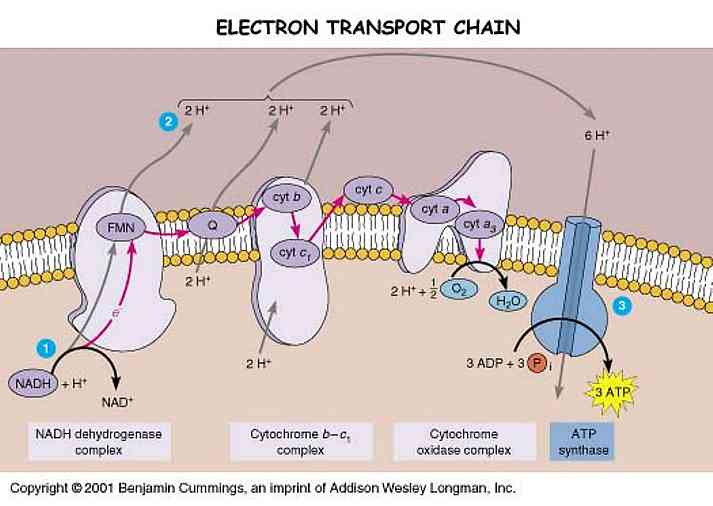
      The potential energy stored in the form of an H+ gradient allows the protons to cross the membrane and produce ATP from ADP. Unlike mitochondria, chloroplasts do not need food energy but transform light energy into chemical energy. In chloroplast, ATP is synthesized as H+ is diffused from thylakoid space back to the stroma though ATP synthase complexes. Therefore, ATP is generated in the stroma.
      In conclusion, thylakoid membrane converts light energy to chemical energy stored in ATP and NADPH. Oxygen is a by-product.

About Photosynthesis

      Photosynthesis is carried out by a number of different organisms, which contain the light-absorbing green-colored pigment chlorophyll. There are several types of chlorophyll found, such as chlorophyll α and chlorophyll β. They are both composed of a porphyrin ring (the light-absorbing portion containing a magnesium atom surrounded by a hydrocarbon ring with alternating single and double bonds. The electrons in the alternating single-double bonds absorb light energy and begin the photosynthetic process) and a long hydrocarbon tail (can anchor the chlorophyll molecule in a membrane by associating with the hydrophobic regions of the phospholipid bilayer. Chlorophyll α and chlorophyll β only differ at the positions of –R. Chlorophyll α contains a methyl group and chlorophyll β contains an aldehyde group, the different functional groups may affects the light energy that the molecules can absorb. Chlorophyll α is the primary light-absorbing pigment, transfers the energy of light to the carbon fixation reactions. Chlorophyll β acts as an accessory pigment, absorbing photons that chlorophyll α absorbs poorly.

Cyanobacteria:
      Cyanobacteria were the first organisms to use sunlight to produce oxygen and organic compounds using water and carbon dioxide. In the endosymbiotic theory, an ancestor of cyanobacteria was engulfed by an ancestor of today’s eukaryotic cells. But unlike plant chloroplasts, they contain chlorophyll d.

Stomata:
      Leaf is the photosynthetic organ of plants, and the stomata on the leaves are regulated by the plants to limit water loss and maximize CO2 intake. The size of stomata is controlled by to guard cells. The water follows the diffusion of potassium ions K+, and K+ also associate with the active transport of H+ ions. During the day, sunlight (light energy) activates specific receptors in guard cell membrane, stimulating proton pumps that drive H+ out of cells. This causes K+ move into the cells, and water to follow by osmosis. CO2 that accumulated in the last night also used up and causes osmosis into the cells. The guard cells swell and the stomata open. When there is no enough water, the guard cells are flaccid and the stomata will be closed.

Chloroplast:
      Chloroplast is where the photosynthesis happens. Chloroplasts have two membranes: outer membrane and inner membrane. Within the chloroplast, the semiliquid material is called stroma, and a system of membrane-bound sacs called thylakoids.
 

Light:
      Electromagnetic (EM) radiation: a form of energy that travels at 3X10^8m/s in the form of wave packets called photons. Photons with short wavelength have high energy and those with long wavelengths have low energy.
      Spectroscope: an instrument that separates different wavelengths into an electromagnetic spectrum.
      Photosystems: clusters of photosynthetic pigments embedded in the thylakoid membranes of chloroplasts that absorb light energy and, through light reaction, transfer the energy from photons to ADP, Pi, and NADP+, forming NADPH and ATP.
      Action spectrum: a graph illustrating the effectiveness with which different wavelengths of light promote photosynthesis.
      Absorption spectrum: a graph illustrating the wavelengths of light absorbed by a pigment.

As you can see, the green light is less likely to be absorbed, therefore it it reflected and the leaves show a green color.

Fermentation

      In the aerobic cellular respiration, NAD+ is reduced into NADH used to produce ATP, and then NADH is oxidized back to NAD+ in ETC. These conversions allow the aerobic cellular respiration to continue and will not be blocked by the lack of NAD+ to start. However, in the anaerobic situation, ETC is not allowed to occur since there is no oxygen acts as the final electron acceptor. Therefore, there are two fermentation processes that can transfer the hydrogen atoms of NADH to cerain organic molecules instead of the ETC.

Ethanol fermentation:
      After finished the glycolysis of one molecule of glucose, two pyruvates, two ATP and two NADH are generated. Instead of undergoing pyruvate oxidation, NADH passes its hydrogen atoms to acetaldehyde, a compound formed when a carbon dioxide molecule is removed from a pyruvate by the enzyme pyruvate decarboxylase. 2 ethanol molecules are produced as the final products, and 2 carbon dioxide molecules are released as the byproducts. Since the NADH generated in the glycolysis are all oxidized back to NAD+, there are only two ATP generated in the process.


      Ethanol fermentation usually carried out by yeast, and used to make breads, wine, beer…

Lactate (Lactic Acid) fermentation:
      Another type of fermentation, that happens during strenuous exercise in human and animals, muscle cells respire glucose faster than oxygen can be supplied, and then oxidative respiration slows down and lactate fermentation begins. NADH generated in the glycolysis transfers its hydrogen atoms to pyruvate in the cytoplasm of the cell, regenerating NAD+ and allowing glycolysis to continue. This results the change of pyruvate into lactate. The accumulation of lactate molecules in muscle tissue causes stiffness, soreness, and fatigue.

Cellular Respiration Regulation

     The reactions of aerobic respiration are regulated by various feedback inhibition and product activation loops. Phosphofructokinase is an important control enzyme in the regulation of cellular respiration.

1)    The catalyst phosphofructokinase at the third reaction of glycolysis is inhibited by ATP from the final product, while it is also stimulated by ADP. Less ATP will be produced when ATP levels are high and ADP levels are low, vice versa.

2)   If the first product of the Krebs cycle, citrate, is accumulated in the mitochondria, some will pass into the cytoplasm and inhibit phosphofructokinase in the glycolysis. As citrate is used and deceased in its concentration, phosphofructokinase inhibition will be reduced, and the rate of glycolysis will increase.

3)   When there is a high concentration of NADH, it indicates that the electron transport chains are full of electrons and ATP production is high. In this case, NADH will inhibit pyruvate decarboxylase and reduces the amount of acetyl-CoA in the process of pyruvate oxidation.

2011年4月3日星期日

Cellular Respiration Details

Stage 1: Glycolysis (anaerobic)
Glycolysis is thought to be the earliest form of energy metabolism. It is an anaerobic process takes place in the cytoplasm that produces two 3-carbon pyruvate molecules from a glucose (6-carbon sugar) molecule. There are 10 steps in glycolysis and 4 ATP and 2 NADH molecules are generated whereas 2 ATP molecules are used.
C6H12O6 + 2 NAD+ + 2 ADP + 2 P -----> 2 pyruvic acid, (CH3(C=O) COOH + 2 ATP + 2 NADH + 2 H+
ATP is required at steps 1 and 3. The hydrolysis of ATP to ADP is coupled with these reactions to transfer phosphate to the molecules at steps 1 and 3.  Reactions 6 and 9 are coupled with the formation of ATP. 2 ATP are produced at step 7 and 2 more ATP molecules are produced at step 10. The net production of ATP is 2. Reaction 6 is an oxidation where NAD+ removes 2 protons and 2 electrons to produce NADH and H+, 2 NADH molecules are generated.

Stage 2: Pyruvate Oxidation (aerobic)
      The two pyruvate molecules formed in glycolysis are transported through the two mitochondrial membranes into the matrix. In each pyruvate, one of the three carbon atoms is cleaved off by the enzyme pyruvate dehydrogenase. This carbon atom attached to oxygen and becomes carbon dioxide as the waste product. This process is known as a decarboxylation reaction. Then the other 2 carbon atoms make up an acetyl group and are added to coenzyme A, forming acetyl coenzyme A. A molecule of NADH is produced in this process. If there is a high concentration of ATP then acetyl-CoA is channeled into an anabolic pathway that synthesized lipids as a way of storing large amounts of energy as fat. If the body does need more energy, the two molecules of acetyl-CoA will enter the Krebs cycle where additional free energy transfers occur.

Stage 3: Krebs cycle (aerobic)
      Krebs cycle is an eight-step process, and each step catalyzed by a specific enzyme. It is a cyclic process since the product of step 8 oxaloacetate (4-carbon) is the reactant in step 1, and combines with acetyl-CoA (2-carbon) which from the pyruvate oxidation to form citrate (6-carbon). By the end of the Krebs cycle, the original glucose molecule is entirely consumed. The six carbon atoms leave the process as six low-energy CO2 molecules as metabolic waste.  In each Krebs cycle, 2 CO2 molecules are produced, 3 NADH, 1 FADH2, and 1ATP are generated.
C6H12O6 + 6O<SUB<2< sub> -> 6CO2 + 6H2O + energy (ATP)

know more about Krebs cycle:


Stage 4 and 5: Electron Transport and Chemiosmosis (aerobic)
      NADH and FADH2 transfer the hydrogen atom electrons they carry to a series of compounds, mainly proteins, which are associated with the inner mitochondrial membrane, so called the electron transport chain (ETC).  The components of the ETC are arranged in order of increasing electronegativity. Each component is reduced by accepting two electrons from the component before the chain and oxidized by losing the electrons to the component after it in the chain. Oxygen acts as the final electron acceptor in the ETC and forms water by combining two H+ from the matrix. Since it is an exergonic process, the free energy released in the process is used to move protons from the mitochondrial matrix to the intermembrane space of mitochondria. Each NADH can pump three protons whereas each FADH2 can pump two, because NADH passes its electrons on to the first protein complex, NADH dehydrogenase, and FADH2 transfers its electrons to Q, the second component of the chain. The NADH produced in the glycolysis in the cytoplasm also diffuse through the outer mitochondrial membrane into the intermembrane space, but it needs to go through the shuttles on the inner membrane to pass on to the matrix, since the inner membrane is impermeable to NADH.
Two shuttle systems:
1)    Glycerol-phosphate shuttle, transfers the electrons from NADH to FAD to form FADH2.
2)   Aspartate shuttle, transfers the electrons to NAD+ instead of FAD and forming NADH.
The accumulation of protons in the intermembrane space of the mitochondrion create an electrochemical gradient that stores free energy, since a higher positive charge and a higher concentration of protons in the intermembrane space (H+ reservoir) are caused. The free energy stored in this gradient produces a proton-motive force and moves protons through the ATPase complex; the free energy lost by the gradient is used as chemical potential energy to drive the synthesis of ATP. The result is 2 ATP per FADH2 and three ATP per NADH are formed. This process is known as chemiosmosis. The ATP molecules are finally produced in the mitochondrial matrix.

Cellular Respiration Overview

     
      Cellular respiration is an exergonic process of gaining energy from organic compounds by steps. The process of using oxygen is called aerobic cellular respiration, and the process of not using oxygen is known as anaerobic cellular respiration. Cellular respiration is a complex sequence of chemical reactions, but the overall equation is simple:
C6H12O6 + 6O2 6CO2 + 6H2O + Energy (as ATP)
Three overall goals of the process:
1.     To break the bonds between the six carbon atoms of glucose, resulting in six carbon dioxide molecules.
2.    To move hydrogen atom electrons from glucose to oxygen, forming six water molecules.
3.    To trap as much of the free energy released in the process as possible in the form of ATP.

Four stages of the process:
1.     Glycolysis
2.    Pyruvate oxidation
3.    The Krebs Cycle (citric acid cycle)
4.    Electron transport and chemiosmosis.

There are two methods to generate energy that produced during the cellular respiration:
1.     Substrate-level phosphorylation:
It is a mechanism to form ATP directly in an enzyme-catalyzed reaction by transferring a phosphate group directly to ADP from a phosphate-containing compound (anaerobic).
For each glucose molecule processed, 4 ATP molecules are generated this way in glycolysis and 2 in the Krebs cycle.
2.    Oxidative phosphorylation:
It is a mechanism to form ATP indirectly through a series of enzyme-catalyzed redox reactions involving oxygen as the final electron acceptor (aerobic).
1)    The coenzyme NAD+ accepts two electrons and one proton from a portion of the original glucose molecule, and reducing to NADH. A dehydrogenase enzyme is used to catalyze this reaction.
NAD+ reduction occurs in one reaction of glycolysis, pyruvate oxidation step, and in three reactions of the Krebs cycle.
2)   The coenzyme FAD is also reduced to FADH2 by accepting two hydrogen atoms (two protons and two electrons).
FAD reduction occurs in one of the reactions of the Krebs cycle.
      The reduced coenzymes act as mobile energy carriers within the cell, moving free energy from one place to another and from one molecule to another (a cell transfers free energy from NADH and FADH2 to ATP). It involves the last stage of the cellular respiration process by using of free oxygen molecules: electron transport and chemiosmosis.

2011年3月19日星期六

Enzyme

      Enzymes are protein catalysts. A catalyst is a substance that speeds up a chemical reaction without being consumed in the process, and ready to catalyze the same reaction again. Although high temperatures can increase the reaction rate, proteins are denatured at high temperatures and cannot function properly any more. In order to speed up the chemical reactions in living cells at moderate temperatures, the protein catalyst enzymes are used to lower the activation energy (Ea) barrier. Enzymes may be grouped into complexes or incorporated into membranes, specific organelle, or cytosol within the cell.

enzymes only lower Ea


      The catalyst does not affect the free energy change of a reaction or the position of equilibrium, it can only decrease the potential energy level of the transition date, therefore, allow a greater proportion of colliding reactants to reach the transition state and become products.
      Substrate is the reactant that an enzyme acts on when it catalyzed a chemical reaction, and the location where the substrate binds to an enzyme is called active site. As the substrate enters the active site on the enzyme, its functional groups come close to the functional groups of a number of amino acids, then an interaction formed between these chemical groups and causes the shape of protein changes, this is known as the induced-fit model. Normally a substrate held in active site by hydrogen bonds and ionic bonds, and an enzyme-substrate complex is created. Active site can lower activation energy by acting as a template for substrate orientation, stressing the substrates and stabilizing the transition state, providing a favorable microenvironment, or participating directly in the catalytic reaction. After the reaction finished, the products are released, and the active site is available for new substrate. Different enzymes have different optimal temperatures and PH levels; the activity of enzyme is always affected by these two environmental factors. Some enzymes also require either nonprotein cofactors, such as zinc ions and manganese ions, or organic coenzymes such as NAD+, FAD+, and NADH.
     
       Enzyme-catalyzed reactions can be saturated, which means every enzyme is used to catalyze the reaction, and no free enzymes are available for more substrates. Therefore, a catalyzed reaction proceeds cannot increase indefinitely by increasing the concentration of the substrate.
     
      Regulation of enzyme catalyzed reaction:
Enzyme inhibition:
      Competitive inhibitors are substances that compete with the substrate for an enzyme’s active site. This process is reversible and can be overcome by increasing the concentration of the substrate. Noncompetitive inhibitors are substances that attach to a binding site on an enzyme other than the active site, causing a change in the enzyme’s shape and a loss of affinity for its substrate. Allosteric inhibitor is an example of noncompetitive inhibitor, a substance that binds to an allosteric site where is a receptor site and some distance from the active site on an enzyme, and stabilized the inactive form of the enzyme. Feedback inhibition is a method to control metabolic pathways allosterically, in which a product formed later in a sequence of reactions allosterically inhibits an enzyme that catalyzed a reaction occurring earlier in the process.

feedback inhibition

Enzyme activation:
      Enzymes can also be active by using of activators. Allosteric activator is a substance that binds to an allosteric site on an enzyme and stabilized the protein conformation that keeps all the active sites available to their substrates. Cooperativity is another type of allosteric activation, by binding one substrate molecules to active site of one subunit and locking all subunits in active conformation.

cooperativity


2011年3月15日星期二

Nucleic acid

       Nucleic acids are informational macromolecules. They are used by all organisms to store hereditary information that determines structural and functional characteristics. They are the only molecules in existence that can produce identical copies of themselves.

DNA and RNA are nucleotide polymers. A nucleotide subunit in both molecules contains a nitrogenous base, a five-carbon sugar, and a phosphate group. The only difference between their monomers is DNA contains the sugar deoxyribose, whereas RNA contains ribose.
      
Nitrogenous base in DNA: A, G, C, T

      
Nitrogenous base in RNA: A, G, C, U


C, T, U are single-ringed pyrimidines, while A, and G are larger double-ringed purines.

A single nucleotide polymer is called a strand. Covalent bonds are formed between the phosphate group of one nucleotide and the hydroxyl group attached to the number 3 carbon of the sugar on the adjacent nucleotide. A phosphate diester bond forms as the result of a condensation reaction between two –OH groups.

DNA is double stranded. The two strands are hold together by hydrogen bonds between nitrogenous bases on adjacent strands, and they are said to run antiparallel.
A—T
G—C
RNA molecules coil into a helix, but remain single stranded.


ATP is another type of nucleotide, which is used to drive virtually all the energy-requiring reactions in a cell. The hydrolysis of ATP is usually coupled with an endergonic process that attached the inorganic phosphate group to another molecule directly associated with the work the cell needs to do.

Protein

Proteins are biochemical compounds consisting of one or more polypeptides folded into a fibrous or globular form. Proteins are coded by the genetic information on DNA.

There are 7 classes of protein:
Ø  Structural proteins (function in the cell membrane, muscle tissue, and the silk of spiders, fibers, and mammal hair.)
Ø  Contractile proteins (work with structural elements, provide muscular movement.)
Ø  Storage proteins (such as albumin, the main substance of egg white.)
Ø  Defensive proteins (antibodies)
Ø  Transport proteins (hemoglobin, which transport oxygen from lungs to other parts of the body.)
Ø  Enzymes (the most important class, promote and regulate the chemical reactions in the cell.)
Ø  Hormones (can act as chemical messengers such as insulin.)

Proteins are amino acid polymers folded into specific three-dimensional shapes. The functions are determined by the specific structural characteristics. An amino acid is an organic molecule, including a central carbon atom which is attached by an amino group, a carboxyl group, an H atom and a variable group of atoms called a side chain (R). There are 20 different amino acids which are different in their side chains.

*amino and carboxyl groups are ionized.
*since amino acids contain both acidic (carboxyl) and basic (amino) groups, amino acids are considered amphiprotic.
*amino acids may be polar, nonpolar, or acidic, basic, depending on the nature of their side chains.
*9 amino acids are considered essential: histidine, isoleucine, leucine, lysine, methionine, phenylalanine, threonine, tryptophan, and valine.
      
An amino acid polymer is called a polypeptide. The amide linkage that holds amino acids together in polypeptides are called peptide bonds. Peptide bonds are formed by a condensation reaction between the amino group of one amino acid and the carboxyl group of an adjacent amino acid.
amino_acid_structure_2

1 A.A a monomer;
2 A.As dipeptide (has one bond);
Many A.As polypeptidesprotein molecule.

Primary protein:
       Primary protein is the unique sequence of amino acids in a polypeptide chain. The polypeptide chain will always have an amino at one end, called the amino terminus, and a carboxyl group at the other, called the carboxyl terminus. Each of the amino acids in a polypeptide is referred to as a residue. Primary structure is determined by the nucleotide sequence in a gene in DNA. Changing the sequence by one amino acid could alter the three-dimensional shape to the point that the protein loses function or is rendered useless.

Secondary protein:
Secondary protein contains coils and folds in a polypeptide caused by hydrogen bonds between hydrogen and oxygen atoms near the peptide bonds.
l  α helix: a type of polypeptide secondary structure characterized by a tight coil that is stabilized by hydrogen bonds.
i.e. keratin.
l  β-pleated sheet: polypeptide secondary structured that form between parallel stretched of a polypeptide and are stabilized by hydrogen bonds.
i.e. silk protein.
*α helix is more flexible than β-pleated sheet, and β-pleated sheet structure is stronger.

Tertiary protein:
Supercoiling of a polypeptide that is stabilized by side-chain interactions.
l  Hydrophobic and van der Waals interactions
l  Proline kink
l  Hydrogen bond
l  Disulfide bridge
l  Ionic bond
 

Quandary protein:
Two or more polypeptide subunits forming a functional protein.
i.e. Hemoglobin: composed of four polypeptides in tertiary structure: two identical α-chains and two identical β-chains. Each subunit has a nonproteinaceous heme group containing an iron atom that binds oxygen.

Protein denaturation:
Protein denaturation is a change in the three-dimensional shape of a protein caused by changes in temperature, PH, ionic concentration, or other environmental factors. A denatured protein cannot carry out its biological functions; all their five types of tertiary interactions are disrupted.
Use of protein denaturation:
Dangerous: prolonged fever above 39 could denature critical enzymes in the brain, leading to seizures and possibly death.
Useful: heat can be used to denature the proteins in hair, allowing people to temporarily straighten curly hair or curl straight hair.

2011年2月25日星期五

Lipids and Fats

Lipid:
Lipid molecules are all hydrophobic since they contain fewer polar O-H bonds and more nonpolar C-H bonds. Lipids are used for storing energy and building membranes.

Fats:
Fats are formed by excess carbohydrate and store energy in both animals and plants. The most common fat is called triacylglycerol or triglyceride, which combined with a glycerol molecule and three fatty acids.
*Glycerol – three carbon alcohol.
*Fatty acid – long hydrocarbon chain containing a single carboxyl group.
Fatty acid can be either saturated or unsaturated. Saturated fatty acids only contain single bonds and make these fatty acids fit more closely together. (Like lard and butter.) Unsaturated fatty acids contain double bonds between carbon atoms, and make the fats in a form of liquid. (Like olive oil and sunflower oil.)
When a glycerol molecule reacts with three fatty acids, three –H atoms from the hydroxyl groups combined with three –OH from the carboxyl groups, and form three water molecules. The bonds between a glycerol and fatty acids are called ester bonds.

Phospholipids:
Phospholipids are made of one glycerol molecule, two fatty acids and a polar phosphate group. Since the phospholipids have both polar and nonpolar parts, they can form a bilayer and act as the main components in cell membrane. The hydrophilic head (polar phosphate group) can mix with water whereas the hydrophobic tails (nonpolar fatty acids) only can mix with each other in the center of bilayer.

Sterols:
Sterols contain four fused hydrocarbon rings and few different functional groups. Cholesterol is an important steroid component of cell membranes. Cholesterol is often converted into vitamin D and bile salts.
Waxes:
Waxes are another lipid containing long-chain fatty acids attached to alcohols or carbon rings.

Waxes are usually used to form waterproof coatings
on plants or animals.